 With age comes changes in the brain. Brain volume decline is a natural occurrence that both men and women deal with. Knowing how to protect the health of your brain as you age is important for slowing down brain aging, supporting cognitive function, and keeping certain diseases at bay. Continue reading to learn more about brain volume decline and steps you can take to maintain brain health as you become older.
Total brain volume comprises the supra- and infratentorial volumes of both gray and white matter, but not the ventricles, brainstem, and choroid plexus. Brain volume can be seen as a representation of the composite reserve for cognitive performance. According to a 2016 article in the Journals of Gerontology, total brain volume is an integrated measure of health and may be an independent indicator of mortality risk independent of any one clinical or subclinical disease state.
An assessment of brain volume is done by the subjective estimation of the relative volume of the cerebrospinal fluid spaces (CSF spaces). Brain volumes can be quantified using structural imaging techniques like automated magnetic resonance imaging (MRI) tools. Also, magnetic resonance imaging (MRI) offers excellent levels of image resolution and between-tissue contrast. As a result, it's often the go-to technique for measuring brain volume.
It’s cited in the article "Brains Over Brawn" that the brain volume of a human is roughly 1,300 cubic centimeters. Normally, men have larger overall brain volumes than women, with differences in the volume of several regions. On one hand, men, for instance, tend to have larger volumes in brain regions related to memory and learning. On the other hand, women tended to have larger volumes in areas of the brain linked with emotion and language. Also, studies suggest that the brain volume of men is between 8% and 13% larger than that of women.
In the book, "Discovering the Brain", it’s maintained that 80% of the brain’s total volume is made up of the cerebral cortex and its supporting structures. It occupies the largest surface area of the human brain. The cortex resembles the bark of a tree with its wrinkled, convoluted appearance, and it covers the surface of the cerebral hemispheres.
Intelligence may be affected by brain size. Research has shown that smaller brain size has the potential to cause issues like poor cognitive performance. Still, research suggests that the intelligence of a person is a result of the brain’s structure and not its size. Researchers contend that the organization of the brain and its molecular activity at the communication junctions or synapses determines the intelligence of an individual.
While the relative volume of your cerebrospinal fluid spaces (CSF spaces) can be an indicator of brain volume, age can play a role as well. The age of a person can impact brain volume. In fact, brain volume diminishes throughout adulthood and with particular health conditions, like alcoholism or dementia.
 According to an article in Postgraduate Medical Journal, as you age your brains shrink in volume, especially in the frontal cortex, as your vasculature (vascular system and arrangements of blood vessels) ages and your blood pressure climbs. Consequently, this increases vascular risk factors and potential for vascular events, such as ischaemia and stroke.
When this happens, the white matter in your brain develops lesions. One study in the Journal of Neurology, Neurosurgery & Psychiatry maintains that white matter lesions in elderly non-demented and demented people are linked to degenerative changes of small vessels. In addition, periventricular white matter lesions are linked to cognitive decline.
Furthermore, studies have revealed that the volume of the brain and/or its weight declines with age at a rate of around 5% every decade after age 40, with the potential for the rate of decline increasing with age, especially over age 70. The myelin sheath (which forms around nerves, including those in the brain) of the white matter deteriorates after around the age of 40, even in normal aging. Subsequently, brain shrinkage appears to be connected to neuronal cell death.
Brain atrophy is typically characteristic of loss of brain cells called neurons and brain shrinkage. This shrinkage can lead to decreased total brain volume. On average, you lose between 0.5% – 1% of your brain volume per year. Still, the rate of loss varies from person to person. One study in Archives of Neurology suggests that this decline in brain volumes may be the result of a nonlinear acceleration in rates of atrophy with increasing age. Also, decreased brain volume has been linked to increased ventricle volume and enlarged superficial sulci.
Furthermore, this is a process that can hasten with injury, infection, or neurodegenerative disorders.
As you age, it’s normal for brain volume loss to occur. With increasing age, your brain shrinks, changing at all levels: molecular, cellular, structural, cognitive, and vascular. So, the older you get, the more brain volume loss usually occurs. Overall brain shrinkage usually starts when a person is in their 30s or 40s. Also, some evidence suggests that the rate of shrinkage increases after the age of 60 or 70. Furthermore, healthy aging has been linked to a steady volume loss in gray and white matter tissues of the brain.
Brain volume shrinkage can be determined through MRI brain scans. Specifically, this technique can show a reduction in brain volume or an increase in the volume of the brain’s ventricles.
The potential symptoms of brain volume decline may differ from person to person depending on the cause. Some common symptoms include:
As you age, there are certain actions that you can take to proactively support your brain health, help prevent a decrease in brain volume, and by extension, slow or prevent cognitive decline.
 Develop an active lifestyle as you age and ensure that you are moving every day. Establishing an exercise routine can help to increase blood flow to your entire body, particularly your brain. Regular physical exercise is beneficial for reducing stress and depression and enhancing memory.
According to an article in Neurology: Clinical Practice, researchers found that exercising for roughly 52 hours total was associated with improved cognitive performance in older adults with and without cognitive impairment. Particularly, aerobic, strength training, mind–body exercises, or combinations of these exercise modes were especially beneficial.
You may not have known that eating a heart-healthy diet is also good for your brain volume. It’s best to adopt a balanced diet. This should be rich in leafy greens, lean proteins, fresh fruits, and healthy fats, and packed with essential nutrients that the body and brain need.
According to an article in the International Journal of Molecular Sciences, while a dietary supply of macronutrients is essential for human health, micronutrients such as vitamins and minerals are also crucial for maintaining brain health. Also, consuming a diet rich in antioxidants and anti-inflammatory components present in fruits, nuts, vegetables, and fish is important. They could potentially minimize age-related cognitive decline and the risk of developing various neurodegenerative diseases.
As you age, you have to be more conscientious about how foods that you eat could impact your brain health. Avoiding an excess of alcohol, salts, sugars, and unhealthy fats is important. Overusing alcohol, for example, can cause memory problems and confusion.
Mental activity should be consistent in order to keep your mind sharp. This also helps to keep your memory and thought processing in tip-top shape. As such, stay mentally active with activities such as reading, playing word games, or learning how to play an instrument. A 2021 study in International Psychogeriatrics suggests that frequent reading activities are associated with a reduced risk of cognitive decline for older adults in the long term.
Maintaining a social life is good for your brain health, by keeping stress and depressive feelings at bay. Having strong bonds with family and friends helps you to stay connected and stay in touch. Based on findings in a 2020 study in Innovation in Ageing, social activities have a significant direct effect on cognitive functioning. Specifically, being socially active via better mental health and physical activities was connected to better future cognitive function in older individuals.
If you care to keep your brain healthy and you are a smoker, quit the habit. Cigarette smoking can wreak havoc not only on your lungs, but it may also contribute to cognitive decline. Researchers found that there is a negative association between cigarette smoking and cortical thickness and cognitive ability. Cigarette smoking causes thinning of the cortex and can lead to cognitive impairment.
Furthermore, quitting smoking can help to bring about positive structural changes in the brain’s cortex. In fact, cortical thickness recovery can occur in as little as a few weeks of quitting according to the findings.
 According to an article in the Journal of Alzheimer's Disease, physical exercise has been shown to increase brain volume and improve cognition in elderly individuals without dementia. Specifically, Tai Chi showed significant increases in total brain volume over the intervention period.
Still, other studies show that moderate to vigorous intensity exercise and higher aerobic fitness can positively impact whole brain volume and gray matter volume. Also, exercise has been shown to effectively increase whole brain volume and gray matter volume in older adults.
Furthermore, other studies suggest that high-quality omega-3 fatty acids circulating in the body are associated with bigger brains. Specifically, one study’s findings revealed that women with twice the levels of omega-3s, DHA and EPA, had roughly 7% larger brain volume compared to women with low levels.
One study shows how brain volume changes in Parkinson's patients. Over time, certain regions of the brain shrink according to recent findings presented in Cortex by researchers of the Human Brain Project (HBP). They indicated that, in Parkinson’s disease, the volumes of particular brain regions decrease over a long period of time. This shrinkage occurs in a specific pattern that’s connected with clinical symptoms.
The new study analyzed the changes in brain volumes in 37 Parkinson's patients and 27 controls using magnetic resonance imaging (MRI). During the initial stage of the study, the researchers found that the volumes of several brain regions were smaller in the Parkinson's disease patients than in the control group. However, it appeared that some enlarged regions were due to compensatory effects.
Over time, the brain volumes of the Parkinson's disease patients declined almost twice as rapidly as those of the control group. In particular, the grey matter showed a significant decline. Also, the volume decrease was quite remarkable in the temporal and occipital lobes.
Furthermore, the team analyzed the brain changes over time using neuroanatomical atlases. It revealed a specific regional pattern of volume changes in Parkinson's disease patients that differed from that which is associated with healthy aging. In fact, the volume decreases of the amygdala and basal forebrain in Parkinson's patients was linked to worsening Parkinson’s symptoms.
Brain volume loss is also a critical feature of multiple sclerosis (MS). A recent study shows that brain volume changes may be a key indicator of MS progression. Specifically, the rate of brain shrinkage may be predictive of disease progression across all stages of MS. While brain shrinkage is a normal part of aging, the rate of brain volume loss may speed up in people with MS. It could be above the average 0.5% – 1% per year.
The changes in brain volume may be an accurate measure of neurodegeneration and tissue damage. Also, cognitive impairment in MS is also linked to brain volume loss as well. As such, brain shrinkage assessment is an important biomarker in MS.
A study published in Annals of Clinical and Translational Neurology analyzed the data of 487 people with primary progressive MS. It included multiple MRI images of their brains and clinical observations. The researchers looked at the rate of brain loss and discovered that a relationship exists between the level of brain volume loss and the progression of the disease. Those with the greatest loss of brain volume had more marked disability and disease progression.
 Brain volumes have been linked to clinical features of attention deficit hyperactivity disorder (ADHD). Researchers found that overall brain volume and several regional volumes were smaller in people with ADHD. Smaller volumes of caudate, cerebellum, and frontal and temporal gray matter are linked to greater ADHD symptom severity.
One study found that individuals diagnosed with ADHD have slightly smaller overall brain volume than non-ADHD people. The results from the study confirmed that individuals with ADHD have differences in their brain structure according to researchers. The findings suggest that ADHD is a disorder of the brain with significant effects on the volumes of subcortical nuclei.
Chronic stress plays a major role in the shrinkage of a particularly critical part of the brain. Adrenal Fatigue Syndrome (AFS) is the non-Addison's form of adrenal dysfunction, where the body's stress response cannot keep up with life's chronic stressors. It puts excess demands on the adrenal glands to secrete cortisol to combat the stress. Eventually, the adrenal glands are depleted. Symptoms emerge at this point.
The effects of chronic stress burden the autonomic nervous system of the Neuroaffect Circuit, which is also comprised of the brain and the microbiome. Stress affects brain functioning and has the potential to bring on significant physical changes to the brain. Shrinking of the medial prefrontal cortex and a loss of gray matter is possible under stress.
Furthermore, too little cortisol means that there isn’t enough of the hormone to counter stress. As such, its effect on the pre-frontal cortex increases. The neuroaffect system is regulated by the brain. In particular, it controls the very part of the brain that has the potential to shrink when you experience stress.
As the brain ages, physical and mental functions become impaired. This natural progression cannot be stopped, but there are certain lifestyle changes that you can adopt to slow the process. Adding brain-boosting supplements to your daily diet can also help with the maintenance of brain health. With the right supplements, you can support and nourish your brain for longevity.
The best supplements to support brain health include the following:
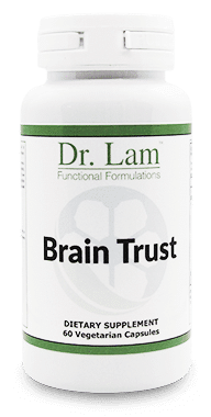
You can also try Dr. Lam’s Brain Trust supplement, which provides maximum support and nourishment to cerebral functions. Its components, Huperzine A and RoseOx®, are known for their neuroprotective and antioxidant activity properties respectively.
However, make sure to talk to a doctor before taking any new supplements to be sure that it doesn't interact with any other medication, supplements, or conditions you may have.
Our brain volume declines with age, and this can have a significant impact on your health. This natural process can be hastened, especially with the presence of certain health conditions. As such, it’s essential that you take the necessary steps to protect your brain as you age. Adopting a healthier lifestyle that includes a healthy balanced diet, regular exercise, hobbies, and stimulating social activities can help.
If you think that your brain may be slowing down and are unsure about your symptoms, get a proper assessment for an accurate determination of the health of your brain. Visit your doctor to know if your symptoms are indicative of brain volume decline.
If you think that you are experiencing symptoms of brain shrinkage and would like to learn about natural ways to remedy them, the team at Dr. Lam Coaching can help. We offer a free** no-obligation phone consultation at +1 (626) 571-1234 where we will privately discuss your health concerns and various options. You can also send us a question through our Ask The Doctor system by clicking here.

Amplify your mind's potential with Brain Trust!
Ackerman S. Discovering the Brain. Washington (DC): National Academies Press (US); 1992. 2, Major Structures and Functions of the Brain. Available from: https://www.ncbi.nlm.nih.gov/books/NBK234157/
Chang, Yu-Hung et al. “Reading activity prevents long-term decline in cognitive function in older people: evidence from a 14-year longitudinal study.” International psychogeriatrics vol. 33,1 (2021): 63-74. doi:10.1017/S1041610220000812
Cohn-Schwartz, Ella. “Pathways From Social Activities to Cognitive Functioning: The Role of Physical Activity and Mental Health.” Innovation in Aging vol. 4,3 igaa015. 30 Jun. 2020, doi:10.1093/geroni/igaa015
de Leeuw, F E et al. “Prevalence of cerebral white matter lesions in elderly people: a population based magnetic resonance imaging study. The Rotterdam Scan Study.” Journal of Neurology, Neurosurgery, and Psychiatry vol. 70,1 (2001): 9-14. doi:10.1136/jnnp.70.1.9
Gomes-Osman, Joyce et al. “Exercise for cognitive brain health in aging: A systematic review for an evaluation of dose.” Neurology. Clinical practice vol. 8,3 (2018): 257-265. doi:10.1212/CPJ.0000000000000460 https://pubmed.ncbi.nlm.nih.gov/30105166/
Halszka Glowacka. (2015, Oct 01). Brains over Brawn. Retrieved June 16, 2023, from https://askananthropologist.asu.edu/stories/brains-over-brawn
Kijonka, Marek et al. “Whole Brain and Cranial Size Adjustments in Volumetric Brain Analyses of Sex- and Age-Related Trends.” Frontiers in Neuroscience vol. 14 278. 3 Apr. 2020, doi:10.3389/fnins.2020.00278
Karama, S, et al. "Cigarette Smoking and Thinning of the Brain'S Cortex." Molecular Psychiatry, vol. 20, no. 6, 2015, pp. 778-785, https://doi.org/10.1038/mp.2014.187. Accessed 20 Jun. 2023.
Koblinsky, Noah D et al. “Household physical activity is positively associated with gray matter volume in older adults.” BMC geriatrics vol. 21,1 104. 5 Feb. 2021, doi:10.1186/s12877-021-02054-8
Melzer, T. M., Manosso, L. M., Gil-Mohapel, J., & Brocardo, P. S. (2021). In Pursuit of Healthy Aging: Effects of Nutrition on Brain Function. International Journal of Molecular Sciences, 22(9). https://doi.org/10.3390/ijms22095026
Miller, David H et al. “Brain atrophy and disability worsening in primary progressive multiple sclerosis: insights from the INFORMS study.” Annals of Clinical and Translational Neurology vol. 5,3 346-356. 30 Jan. 2018, doi:10.1002/acn3.534
Mortimer, James, et al. "Changes in Brain Volume and Cognition in a Randomized Trial of Exercise and Social Interaction in a Community-Based Sample of Non-Demented Chinese Elders." Journal of Alzheimer'S Disease : JAD, vol. 30, no. 4, 2011, p. 757, https://doi.org/10.3233/JAD-2012-120079. Accessed 20 Jun. 2023.
Peters, R. (2006). Ageing and the brain. Postgraduate Medical Journal, 82(964), 84-88. https://doi.org/10.1136/pgmj.2005.036665
Pieperhoff, P., et al. (2022) Regional changes of brain structure during progression of idiopathic Parkinson's disease – A longitudinal study using deformation based morphometry. Cortex. doi.org/10.1016/j.cortex.2022.03.009.
Pottala, James V et al. “Higher RBC EPA + DHA corresponds with larger total brain and hippocampal volumes: WHIMS-MRI study.” Neurology vol. 82,5 (2014): 435-42. doi:10.1212/WNL.0000000000000080
Scahill, Rachael I et al. “A longitudinal study of brain volume changes in normal aging using serial registered magnetic resonance imaging.” Archives of Neurology vol. 60,7 (2003): 989-94. doi:10.1001/archneur.60.7.989
Van Elderen, Saskia S G C et al. “Brain Volume as an Integrated Marker for the Risk of Death in a Community-Based Sample: Age Gene/Environment Susceptibility--Reykjavik Study.” The Journal of Gerontology. Series A, Biological sciences and medical sciences vol. 71,1 (2016): 131-7. doi:10.1093/gerona/glu192 https://academic.oup.com/biomedgerontology/article/71/1/131/2614153
Yale University. (2012, August 12). How stress and depression can shrink the brain. ScienceDaily. Retrieved June 14, 2023, from https://www.sciencedaily.com/releases/2012/08/120812151659.htm
Total brain volume comprises supra- and infratentorial volumes of both gray and white matter and can be seen as a representation of the composite reserve for cognitive performance. You can maintain brain health by eating healthy, exercising regularly, staying mentally active, quitting smoking, and maintaining a social life.